-
Question 1
Incorrect
-
A 30-year-old woman is receiving IV co-trimoxazole in the hospital. She initially showed signs of improvement, but now she has developed a fever, joint pain, oral and genital ulcers, and target-like lesions on her palms and soles that have spread to her face and trunk. The lesions have become tender and blistered, and lateral pressure causes erosions. The vancomycin has been discontinued.
What is the most appropriate next step in managing this patient?Your Answer: Withdrawal of suspected drug
Correct Answer: Intravenous fluid supplementation
Explanation:Treatment Options for Stevens-Johnson Syndrome
Stevens-Johnson syndrome (SJS) is often triggered by medications, and prompt withdrawal of the suspected drug is crucial for improving prognosis. The most commonly associated drugs include allopurinol, aromatic anticonvulsants, antibacterial sulfonamides, and oxicam NSAIDs. Supportive care for SJS is similar to that for major burns and wound care, including intravenous fluid supplementation. Analgesia may also be necessary.
Debridement should only be considered for patients with necrotic skin or non-healing wounds. Systemic corticosteroids have not been evaluated in clinical trials, and a systematic review did not suggest improved survival rates. Topical steroids may be prescribed, but stopping the associated drug and providing intravenous hydration are more important. Overall, early diagnosis and withdrawal of suspected drugs are crucial for managing SJS.
-
This question is part of the following fields:
- Dermatology
-
-
Question 2
Incorrect
-
A 35-year-old woman presents to the Genitourinary Clinic with a 4-day history of vulval itching. She denies any fever or lower urinary tract symptoms and reports no vaginal discharge. She had unprotected sex with a new partner three weeks ago. She has no significant medical history and is not taking any medication other than regular folic acid. She has no known allergies. She is currently 14 weeks pregnant.
During the examination, her temperature is 36.5 °C, heart rate is 80 bpm, and blood pressure is 120/70 mmHg. Her abdomen is soft and non-tender. Multiple flesh-colored papules are visible around the vulva, with the largest measuring 4 mm in diameter. Some of the papules have an irregular border with mild bleeding.
What is the most appropriate next step in managing this patient?Your Answer: Topical podophyllotoxin
Correct Answer: Cryotherapy
Explanation:Treatment Options for Anogenital Warts in Pregnancy
Anogenital warts, caused by human papillomavirus (HPV), can be a concern during pregnancy as they can be transmitted to the newborn. Cryotherapy is the recommended treatment for anogenital warts during pregnancy, as per British Association for Sexual Health and HIV (BASHH) guidelines. This treatment involves freezing the warts and may need to be repeated every 1-3 weeks until the warts resolve. Topical imiquimod is another treatment option, but there is limited data on its safety during pregnancy. Oral aciclovir is used to treat herpes virus, which presents with painful vesicles rather than fleshy protuberances. Topical clotrimazole is used to treat thrush, which presents with severe itch and a thick, white discharge. Topical podophyllotoxin is the first-line treatment for anogenital warts, but it is teratogenic and not recommended during pregnancy.
-
This question is part of the following fields:
- Dermatology
-
-
Question 3
Correct
-
A 16-year-old girl comes to your clinic with a complaint of a rash that has been present for a year. She reports that the rash appears on her face and dorsal arms after sun exposure. The rash consists of small red bumps that disappear after a few days without leaving any scars. The patient is healthy otherwise, and her laboratory tests are normal. What is the most likely diagnosis?
Your Answer: Polymorphous light eruption
Explanation:Polymorphous Light Eruption and Similar Skin Conditions
Polymorphous light eruption is a skin condition that commonly affects young females. It can manifest in various forms such as macules, papules, patches, or plaques. However, the rash usually appears the same whenever it recurs. It typically occurs a few hours after sun exposure and resolves within a few days without leaving scars.
Acute lupus erythematosus may present with a similar rash to polymorphous light eruption, but patients will usually experience other symptoms such as joint pains, oral ulcers, and hair loss. They also tend to have positive anti-nuclear antibodies. On the other hand, discoid lupus erythematosus presents with chronic scaly, erythematous-to-hyperpigmented papules and plaques that leave residual scars and pigmentation. This condition is worsened by sun exposure.
Solar urticaria, on the other hand, presents with urticaria upon sun exposure, which usually resolves within a few hours instead of a few days. Meanwhile, patients with xeroderma pigmentosum have an extreme sensitivity to the sun from a very young age. They also present with multiple solar lentigines, nevi, and malignant skin tumors such as basal cell carcinoma, squamous cell carcinoma, and malignant melanoma.
In summary, while these skin conditions may present with similar rashes, they have distinct characteristics that differentiate them from each other. It is important to seek medical attention for proper diagnosis and treatment.
-
This question is part of the following fields:
- Dermatology
-
-
Question 4
Incorrect
-
A 25-year-old female patient with psoriasis is seen at your Dermatology Clinic. She has tried various creams and undergone multiple courses of ultraviolet B (UVB) therapy, but her condition persists. During the consultation, she expresses her frustration and tearfully explains how psoriasis is affecting her social life and self-esteem. She is hesitant to consider systemic therapies that may compromise her immune system. You suggest acitretin as a potential treatment option and inquire about her plans for having children. She responds that she is currently in a relationship but has no immediate plans for starting a family. What is the recommended duration of contraception after taking acitretin?
Your Answer: 6 Months
Correct Answer: 3 Years
Explanation:Contraceptive Precautions for Medications Affecting Pregnancy
When it comes to medications that may impact pregnancy, it is important to take contraceptive precautions to avoid unwanted conception. For acitretin, the manufacturer’s guidelines state that pregnancy must be reliably prevented for 4 weeks before, during, and for 3 years after completing therapy. Isotretinoin and alitretinoin should be discontinued at least 1 month before pregnancy. One week and two weeks are considered too short of a period to avoid conception, as they are less than the duration of a single menstrual cycle. On the other hand, six months is the recommended period of pregnancy avoidance after methotrexate therapy. It is also important to note that males who are sexually active and starting acitretin therapy should also receive advice on contraception.
-
This question is part of the following fields:
- Dermatology
-
-
Question 5
Incorrect
-
A 40-year-old woman with a history of psoriasis visits your clinic with complaints of a severe flare-up. She appears distressed and reveals that her condition is significantly impacting her daily life. The patient is currently taking methotrexate and folic acid for long-term maintenance, following an unsuccessful attempt at phototherapy and an apparent allergic reaction to cyclosporin. What would be your next course of action in managing her condition?
Your Answer: Non-steroidal anti-inflammatory drugs (NSAIDs)
Correct Answer: Biologic immune modifying agent
Explanation:Psoriasis can have a significant impact on a patient’s mental health, and controlling the disease is crucial. Treatment options include topical and systemic methods, with biological immune modifying agents being effective for moderate to severe cases. Topical treatments like coal tar and steroids can help with mild to moderate cases, while systemic treatments like methotrexate and cyclosporin can be used for more severe cases. NSAIDs and topical coal tar are not effective if methotrexate has failed, and steroids should only be used for short-term treatment.
-
This question is part of the following fields:
- Dermatology
-
-
Question 6
Incorrect
-
A 28-year-old man arrives at the emergency department complaining of a fever and a painful rash. He had recently been prescribed oral amoxicillin by his GP to treat a chest infection. Although his cough had improved, he noticed the rash shortly after. He then experienced joint pain and a general feeling of being unwell. Upon examination, he has a maculopapular rash with target lesions, blistering lesions, and mucosal erosions, including in his oral cavity. His chest appears normal. What timing of exposure is most indicative of Stevens-Johnson syndrome?
Your Answer: Within minutes
Correct Answer: Within 1-4 weeks
Explanation:The expected timeframe for the development of Stevens-Johnson syndrome (SJS) after taking amoxicillin is within 1-4 weeks, with an average onset of 14 days. This patient’s symptoms, including a maculopapular rash with blistering lesions, oral involvement, fever, and joint pain, are consistent with SJS. While secondary exposure can sometimes lead to an earlier reaction within 48 hours, this is less common. It is also possible that the symptoms are related to a Mycoplasma or Streptococcal infection. Other drug reactions, such as drug reaction with eosinophilia and systemic symptoms, typically occur within 2-6 weeks, while acute generalised exanthematous pustulosis usually develops within 3-5 days.
Understanding Stevens-Johnson Syndrome
Stevens-Johnson syndrome is a severe reaction that affects the skin and mucosa, and is usually caused by a drug reaction. It was previously thought to be a severe form of erythema multiforme, but is now considered a separate entity. The condition can be caused by drugs such as penicillin, sulphonamides, lamotrigine, carbamazepine, phenytoin, allopurinol, NSAIDs, and oral contraceptive pills.
The rash associated with Stevens-Johnson syndrome is typically maculopapular, with target lesions being characteristic. It may develop into vesicles or bullae, and the Nikolsky sign is positive in erythematous areas, meaning that blisters and erosions appear when the skin is rubbed gently. Mucosal involvement and systemic symptoms such as fever and arthralgia may also occur.
Hospital admission is required for supportive treatment of Stevens-Johnson syndrome. It is important to identify and discontinue the causative drug, and to manage the symptoms of the condition. With prompt and appropriate treatment, the prognosis for Stevens-Johnson syndrome can be good.
-
This question is part of the following fields:
- Dermatology
-
-
Question 7
Incorrect
-
A 35-year-old man presents to the cardiology clinic with bilateral foot pain that has been ongoing for 7 months. Initially, the pain only occurred at the end of his daily commute but has since progressed to being provoked by short distances and occasionally occurring at rest. He also experiences pain in his right wrist. He has no significant medical history but is a current smoker with a 20 pack year history. On examination, his feet are smooth and hairless, and there is difficulty feeling the right dorsalis pedis with cold and dusky toes. Urgent angiography reveals distal stenosis in both limbs with collateral formation. Blood tests are unremarkable except for an elevated CRP. What is the most likely diagnosis?
Your Answer: Microscopic polyangiitis
Correct Answer: Thromboangiitis obliterans
Explanation:Upon examination of the patient, it is evident that they are displaying symptoms of arterial insufficiency. However, their age, lack of vascular risk factors, and upper limb involvement suggest that this may not be a straightforward case of peripheral vascular disease.
There are two vasculitic disorders that could be causing the patient’s symptoms: microscopic polyangiitis and polyarteritis nodosa. Both of these conditions are characterized by constitutional symptoms such as weight loss, fever, anorexia, and fatigue. Renal involvement and skin rash are common, and there is often an association with p-ANCA (MPO) antibodies and an elevation of inflammatory markers.
Takayasu arteritis is a large vessel vasculitis that primarily affects the aorta and its branches. It is more prevalent in females and would not typically cause the isolated small and medium vessel stenoses described above.
Thromboangiitis obliterans is an inflammatory disorder that affects the small to medium-sized arteries and superficial veins in the distal extremities. Peripheral limb ischemia is caused by segmental arterial inflammation and thrombus formation, and the disease often progresses aggressively. Although the etiology is unclear, the condition is strongly linked to smoking, and disease progression is closely tied to continued tobacco use. It is most commonly found in men aged 20-40.
To diagnose the patient’s condition, a thorough history and examination, angiographic findings, and the exclusion of proximal emboli and vasculitic disorders are necessary. Angiography typically reveals segmental occlusions of the distal arteries in the hands and feet, corkscrew collateral vessels, and a lack of widespread atherosclerotic disease. Therefore, thromboangiitis obliterans is the most likely diagnosis.
Overview of Vasculitides
Vasculitides refer to a group of disorders characterized by inflammation of blood vessels. They can affect vessels of different sizes, including large, medium, and small vessels. Large vessel vasculitides include temporal arteritis and Takayasu arteritis, while medium vessel vasculitides include polyarteritis nodosa and Kawasaki disease. Small vessel vasculitides are further divided into ANCA-associated vasculitides, granulomatosis with polyangiitis (Wegener’s granulomatosis), eosinophilic granulomatosis with polyangiitis (Churg-Strauss syndrome), microscopic polyangiitis, immune complex small-vessel vasculitis, Henoch-Schonlein purpura, Goodpasture’s syndrome (anti-glomerular basement membrane disease), cryoglobulinaemic vasculitis, and hypocomplementemic urticarial vasculitis (anti-C1q vasculitis).
In summary, vasculitides are a group of disorders that involve inflammation of blood vessels of different sizes. The classification of vasculitides is based on the size of the affected vessels, with large, medium, and small vessel vasculitides being the main categories. Each type of vasculitis has its own unique clinical features, and prompt diagnosis and treatment are essential to prevent complications and improve outcomes.
-
This question is part of the following fields:
- Dermatology
-
-
Question 8
Incorrect
-
A 20-year-old man with a history of learning difficulties and eczema presents to the dermatology clinic with his parents. Despite using E45 cream three times a day, his eczema is difficult to control and has had several episodes of infection in the past. Currently, there is no sign of infection, but his flexural folds are erythematous with clear discharge. His parents are seeking advice on additional measures to manage his eczema.
Your Answer: Oral steroids
Correct Answer: Bandaging after emollient use
Explanation:To control itching in eczema and prevent further scratching, bandaging is a recommended solution. Bandaging can be used in combination with emollients and, if necessary, topical corticosteroids for non-infected patients with weeping or lichenified skin. For this patient with weeping skin, bandaging can help absorb the discharge and enhance the emollient effect. Severe cases or those that do not respond to topical treatment may require oral steroids or topical immunosuppressant therapy. While urea-containing topical agents can moisturize and relieve itching, they can also worsen inflammation and are not suitable for this patient. Long-term antibiotics are not typically used for eczema.
Eczema is a skin condition that can be severe in some cases. Certain factors can indicate a higher likelihood of severe disease, such as onset at a young age, a history of severe disease in childhood, and the presence of asthma or hay fever. Additionally, having a small family size and high levels of IgE in the blood can also be prognostic markers for severe eczema.
To manage eczema, there are several treatment options available. Emollients can help to moisturize and soothe the skin, while topical steroids can reduce inflammation and itching. UV radiation can also be used to treat eczema, as well as immunosuppressants like ciclosporin, antihistamines, and azathioprine. It is important to work with a healthcare provider to determine the best course of treatment for each individual case of eczema.
-
This question is part of the following fields:
- Dermatology
-
-
Question 9
Incorrect
-
You are evaluating a 60-year-old man who has just commenced treatment for urological cancer with Nivolumab (Opdivo), a type of checkpoint inhibitor. He has been receiving treatment for a month and has not experienced any adverse effects thus far. He is curious about what he can anticipate from this therapy.
What is the most prevalent side effect that this patient is likely to encounter?Your Answer: Depression and anxiety
Correct Answer: Dry itchy skin and rashes
Explanation:Immune checkpoint inhibitors are a type of immunotherapy used to treat certain types of cancer. Their side effects are inflammatory and autoimmune in nature, with dry itchy skin and rashes being the most common. Other side effects include nausea, vomiting, decreased appetite, diarrhea, tiredness, fatigue, shortness of breath, and dry cough.
Understanding Immune Checkpoint Inhibitors
Immune checkpoint inhibitors are a type of immunotherapy that is becoming increasingly popular in the treatment of certain types of cancer. Unlike traditional therapies such as chemotherapy, these targeted treatments work by harnessing the body’s natural anti-cancer immune response. They boost the immune system’s ability to attack and destroy cancer cells, rather than directly affecting their growth and proliferation.
T-cells are an essential part of our immune system that helps destroy cancer cells. However, some cancer cells produce high levels of proteins that turn T-cells off. Checkpoint inhibitors block this process and reactivate and increase the body’s T-cell population, enhancing the immune system’s ability to recognize and fight cancer cells.
There are different types of immune checkpoint inhibitors, including Ipilimumab, Nivolumab, Pembrolizumab, Atezolizumab, Avelumab, and Durvalumab. These drugs block specific proteins found on T-cells and cancer cells, such as CTLA-4, PD-1, and PD-L1. They are administered by injection or intravenous infusion and can be given as a single-agent treatment or combined with chemotherapy or each other.
However, the mechanism of action of these drugs can result in side effects termed ‘Immune-related adverse events’ that are inflammatory and autoimmune in nature. This is because all immune cells are boosted by these drugs, not just the ones that target cancer. The overactive T-cells can produce side effects such as dry, itchy skin and rashes, nausea and vomiting, decreased appetite, diarrhea, tiredness and fatigue, shortness of breath, and a dry cough. Management of such side effects reflects the inflammatory nature, often involving corticosteroids. It is important to monitor liver, kidney, and thyroid function as these drugs can affect these organs.
In conclusion, the early success of immune checkpoint inhibitors in solid tumors has generated tremendous interest in further developing and exploring these strategies across the oncology disease spectrum. Ongoing testing in clinical trials creates new hope for patients affected by other types of disease.
-
This question is part of the following fields:
- Dermatology
-
-
Question 10
Incorrect
-
How would you describe the multiple, small, raised, slightly dome-shaped lesions containing purulent material observed during a skin examination of a patient?
Your Answer: Fissures
Correct Answer: Pustules
Explanation:Skin Lesions: Pustules, Bullae, Fissures, Telangiectasias, and Ulcers
Pustules, bullae, fissures, telangiectasias, and ulcers are all types of skin lesions that can occur for various reasons. Pustules are small bumps on the skin that contain pus or necrotic inflammatory cells. Bullae, on the other hand, are larger fluid-filled blisters that can be caused by burns, infections, or autoimmune disorders. Fissures are deep cracks in the skin that can be painful and are often caused by dryness or trauma. Telangiectasias are visible collections of enlarged capillaries that can appear on the skin or mucous membranes. Finally, ulcers are open sores that can occur when the skin is damaged or breaks down, often due to infection or poor circulation.
-
This question is part of the following fields:
- Dermatology
-
-
Question 11
Incorrect
-
A 42-year-old man visits the Dermatology Clinic with a poorly defined, pearly nodule measuring 0.8 cm in diameter. The lesion is located in the medial canthus, 7 mm from his right lower eyelid, and is accompanied by arborising telangiectasia. What is the most effective treatment option to prevent recurrence and achieve optimal cosmetic results?
Your Answer: Curette and cautery
Correct Answer: Mohs micrographic surgery
Explanation:Basal cell carcinoma (BCC) is a common type of skin cancer, and there are several treatment options available. Mohs micrographic surgery is a highly effective technique that is indicated for recurrent or incompletely excised BCC, primary BCC with indistinct borders, lesions located in or near high-risk areas, and aggressive clinical evolution or histological subtype. Radiotherapy may be used in patients who are not suitable for surgery. Excision with a 5 mm margin and flap repair is an option, but it carries the risk of a poor cosmetic result. Curette and cautery is suitable for small nodular or superficial lesions. Vismodegib is a medication that is used for metastatic BCCs, those that have relapsed after surgery, and those that are not amenable to surgery or radiotherapy treatment. It works by blocking the hedgehog signaling pathway. It is important to discuss the various treatment options with a healthcare provider to determine the best course of action for each individual case.
-
This question is part of the following fields:
- Dermatology
-
-
Question 12
Incorrect
-
A 22-year-old man presents to the dermatology clinic with a severe worsening of his eczema symptoms. Despite using emollients and moderate to severe strength topical steroids, he has experienced a severe flare that did not respond to a one-week trial of oral prednisolone. He is currently experiencing severe pruritus and skin irritation, with evidence of scratch marks on the flexural surfaces of his arms and legs. What non-steroidal treatment options may be effective in controlling his flare?
Your Answer: IV tacrolimus
Correct Answer: Topical tacrolimus
Explanation:When emollients and strong steroid therapy fail to treat severe eczema, dermatologists may turn to calcineurin inhibitors like tacrolimus as an alternative to topical steroids. It’s important to note that oral and IV tacrolimus are typically used for preventing and treating organ rejection, not eczema. Salicylic acid is specifically used for scales in psoriasis, and alitretinoin is reserved for severe cases of refractory hand eczema.
Eczema is a skin condition that can be severe in some cases. Certain factors can indicate a higher likelihood of severe disease, such as onset at a young age, a history of severe disease in childhood, and the presence of asthma or hay fever. Additionally, having a small family size and high levels of IgE in the blood can also be prognostic markers for severe eczema.
To manage eczema, there are several treatment options available. Emollients can help to moisturize and soothe the skin, while topical steroids can reduce inflammation and itching. UV radiation can also be used to treat eczema, as well as immunosuppressants like ciclosporin, antihistamines, and azathioprine. It is important to work with a healthcare provider to determine the best course of treatment for each individual case of eczema.
-
This question is part of the following fields:
- Dermatology
-
-
Question 13
Correct
-
An 80-year-old woman comes to the Emergency Department (ED) complaining of occasional pain in her upper left chest. She has had a blistering rash in the area for the past four days, which has been oozing and causing a burning sensation. She had chickenpox as a child.
During the examination, a clearly defined rash is observed, consisting of scabbed and healing blisters that correspond to a single dermatome.
What is the best course of treatment for this patient?Your Answer: Gabapentin
Explanation:Treatment options for Herpes zoster reactivation (shingles)
Herpes zoster reactivation, also known as shingles, can cause painful lesions. Antiviral therapy is only useful if new lesions are appearing at the time of presentation, indicating ongoing viral replication. However, after lesions have encrusted, antiviral therapy is unlikely to be beneficial. Gabapentin is a useful oral agent to block the pain associated with post-herpetic neuralgia. Aciclovir ointment is mostly useful against conjunctival varicella zoster. Oral valciclovir is an alternative option to acyclovir, with good cover against herpes viruses. IV aciclovir is most appropriate for HSV encephalitis. Alternatives to gabapentin include tricyclic anti-depressants and topical capsaicin cream. The most appropriate starting dose of gabapentin is 300 mg daily, with effective doses being from 900 mg upwards, and it can be increased up to 3.6 g/day.
Treatment options for Herpes zoster reactivation (shingles)
-
This question is part of the following fields:
- Dermatology
-
-
Question 14
Incorrect
-
A 25-year-old woman presents to the Dermatology Clinic after returning from a backpacking trip in Southeast Asia. She complains of a persistent and intensely itchy rash on her hands and feet. Upon examination, linear tracks with secondary blistering and crusting are observed, leading to a diagnosis of cutaneous larva migrans. What is the most probable source of infection for this patient?
Your Answer: Fish
Correct Answer: Dog
Explanation:Parasitic Skin Infections: Causes and Interventions
Cutaneous larva migrans is a parasitic skin infection caused by hookworm that primarily affects domestic animals like dogs, cats, and sometimes cattle. The larvae penetrate the animal’s skin and mature in the intestine, laying eggs that are excreted in the faeces. Humans can contract the infection by coming into contact with contaminated soil or sand on beaches. Albendazole and ivermectin are potential interventions for this infection.
Sand fly bites, on the other hand, can lead to the development of cutaneous leishmaniasis, a different parasitic skin infection. Fish tapeworm is another potential problem that can cause intestinal infection from uncooked freshwater fish, such as sushi. Red ant bites may cause allergic dermatitis in rare cases, but it does not lead to the development of cutaneous larva migrans. Finally, turkeys suffer from caecal worms, which carry the protozoan parasite Histomonas, leading to death in the turkeys, but it is not a cause of human disease.
-
This question is part of the following fields:
- Dermatology
-
-
Question 15
Incorrect
-
A 45-year-old African American man presents to the Neurology Clinic with a 7-month history of distal sensory loss and weakness. He has no history of headache or visual changes and his co-ordination remains intact. He denies any constitutional symptoms or recent infections. He has a past medical history of diet-controlled type II diabetes and hypertension, and takes ramipril 5 mg once daily. On examination, he has visible wasting in the intrinsic muscles of his left foot, with a Medical Research Council (MRC) power of 4/5 for ankle dorsiflexion. There is associated loss of temperature and light touch sensation down the posterolateral aspect of his left leg. The remainder of his neurological examination shows no further motor deficits and his cranial nerves are intact. He has multiple hypopigmented patches over the extensor surfaces of his arms and legs, with associated sensory loss for all but deep pressure stimuli. What is the most appropriate investigation to confirm the diagnosis?
Your Answer: Lumbar puncture
Correct Answer: Skin-slit smear
Explanation:Diagnosing Leprosy: The Role of Skin-Slit Smear
Leprosy is a granulomatous disease caused by Mycobacterium leprae that affects the peripheral nerves and skin. The initial symptoms include numbness, loss of temperature, light touch, pain, and deep pressure. Cutaneous lesions appear later in the disease as hypopigmented patches of skin with associated sensory loss. The investigation of choice for diagnosing leprosy is a skin-slit smear. This involves making a slit over a lesion of skin and smearing it onto a glass slide, which is then examined for acid-fast bacilli under microscopy. If detected, the treatment of choice is with dapsone, rifampicin, and clofazimine.
Other diagnostic methods, such as nerve conduction studies, lumbar puncture, viral DNA polymerase chain reaction, and MRI brain, may be useful in investigating underlying neurological illnesses but are not necessary for diagnosing leprosy. Skin-slit smear remains the most effective and reliable method for diagnosing leprosy.
-
This question is part of the following fields:
- Dermatology
-
-
Question 16
Incorrect
-
A 32-year-old man visits his primary care physician complaining of rapidly developing white patches on his skin over the face, trunk, and limbs for the past two months. During the examination, his blood pressure is 110/70 mmHg, and his heart rate is 96 bpm. He experiences postural hypotension. A diffuse swelling is detected in the front of his neck, which moves with swallowing. Upon further investigation, anti-microsomal and anti-thyroglobulin antibodies are found to be positive.
Lab Results Normal Range
Free T3 (fT4) 6.8 pmol/l 11–22 pmol/l
Free T4 (fT3) 2.3 pmol/l 3.5–5 pmol/l
Thyroid-stimulating hormone (TSH) 70 µU/l 0.17–3.2 µU/l
What would be the appropriate initial treatment for this patient?Your Answer: Punch grafting
Correct Answer: Oral betamethasone pulse therapy with NBUVB
Explanation:Management of Unstable and Progressive Vitiligo: Oral Betamethasone Pulse Therapy with NBUVB
Vitiligo is a skin disorder characterized by the loss of melanocytes, resulting in depigmented patches on the skin. In cases of unstable and progressive vitiligo involving a larger body surface area, oral betamethasone pulse therapy with narrow-band ultraviolet B (NBUVB) is the most appropriate choice for management. This therapy involves high-dose betamethasone pulse therapy on weekends for 3-6 months, along with NBUVB phototherapy twice to thrice weekly.
For patients with rapidly progressive vitiligo, initiation of oral betamethasone pulse therapy with NBUVB after sending 0800 serum cortisol is recommended. Surgery, such as punch grafting, is not an option unless the disease has been stable for a year. Monotherapy with NBUVB phototherapy or topical corticosteroids will not be effective enough to control active disease.
It is important to note that patients with autoimmune thyroiditis, which is commonly associated with vitiligo, should not receive thyroxine replacement before ruling out autoimmune adrenalitis. Starting thyroxine replacement before steroid replacement can further catabolize glucocorticoids and potentially precipitate an Addisonian crisis. In patients with postural hypotension, serum cortisol taken at 0800 should be sent to rule out adrenal insufficiency before initiating any thyroid hormone replacement.
-
This question is part of the following fields:
- Dermatology
-
-
Question 17
Correct
-
A 31-year-old man with autism spectrum disorder visits the general medical clinic due to a severe eczema flare that has not been controlled with emollients, strong topical corticosteroids, and non-sedating antihistamines. He has no other medical issues but is anxious about the spreading rash. During the examination, spreading erythema is observed from flexural folds on his arms and legs. What is the best course of treatment to pursue while waiting for a dermatology appointment?
Your Answer: Start prednisolone 30 mg OD for one week
Explanation:For patients experiencing eczema flares, treatment with prednisolone requires several weeks of medication followed by a gradual tapering off to prevent rebound. In this case, the most appropriate course of action would be to try oral prednisolone for a trial period. According to NICE guidelines, the patient should take 30 mg once a day for one week while awaiting a dermatology review. This dosage can be extended as needed, but close monitoring is necessary. This approach is particularly important for patients experiencing severe eczema and psychological distress. Hospitalization is not required unless the patient is unstable or has eczema herpeticum. For weeping or lichenified eczema, bandaging is a more effective treatment option. Urea-containing emollients can also be helpful for pruritic skin that requires greater hydration in the absence of severe inflammation.
Eczema is a skin condition that can be severe in some cases. Certain factors can indicate a higher likelihood of severe disease, such as onset at a young age, a history of severe disease in childhood, and the presence of asthma or hay fever. Additionally, having a small family size and high levels of IgE in the blood can also be prognostic markers for severe eczema.
To manage eczema, there are several treatment options available. Emollients can help to moisturize and soothe the skin, while topical steroids can reduce inflammation and itching. UV radiation can also be used to treat eczema, as well as immunosuppressants like ciclosporin, antihistamines, and azathioprine. It is important to work with a healthcare provider to determine the best course of treatment for each individual case of eczema.
-
This question is part of the following fields:
- Dermatology
-
-
Question 18
Incorrect
-
A 35-year-old man visits the Dermatology Clinic with a complaint of persistent cracking of the skin at the corners of his mouth. He adheres to a strict vegetarian diet.
What is the most probable reason for his skin changes?Your Answer: Vitamin C
Correct Answer: Riboflavin
Explanation:Potential Vitamin Deficiencies in Vegans and Their Consequences
Vegans are aware of the potential vitamin deficiencies that may arise from their diet and are careful to replace animal sources of B vitamins. However, riboflavin deficiency is still a possibility and can cause angular stomatitis. The most appropriate intervention is dietary modification with oral supplementation.
Vitamin C deficiency can lead to scurvy, which manifests as follicular keratosis, coiling of hair, and perifollicular hemorrhages. It also increases the risk of lower limb hemorrhage.
Vitamin A deficiency can impair dark adaptation of vision and cause dry, scaling skin. This can lead to permanent corneal scarring (keratomalacia) and an increased risk of respiratory infection.
Vitamin D deficiency can result in osteomalacia, which causes long bone pain and proximal muscle weakness. It is more common in individuals who follow a vegetarian diet.
Vitamin K deficiency in adults is rare, as it is found in green vegetables. However, it may occur due to malabsorption of fat and can lead to easy bruising and an increased risk of bleeding.
-
This question is part of the following fields:
- Dermatology
-
-
Question 19
Incorrect
-
A 22-year-old woman arrives at the emergency department with a pruritic rash that appeared during a day spent at the beach with friends. She reports experiencing a burning and itching sensation on her upper arms, trunk, and thighs. Upon examination, multiple 3mm pink papules are observed on the affected areas, with her face and hands being spared. What is the probable diagnosis?
Your Answer: Systemic lupus erythematosus
Correct Answer: Polymorphic light eruption
Explanation:Photoallergic contact dermatitis is a reaction that can occur when certain chemicals are applied to the skin and then exposed to sunlight. This reaction can affect any part of the body, including the hands and face.
Xeroderma pigmentosum is a rare genetic disorder that affects the body’s ability to repair DNA damage caused by exposure to ultraviolet rays. This condition can cause extreme sensitivity to sunlight and increase the risk of developing skin cancer. Symptoms typically appear in childhood and include multiple basal cell carcinomas and other types of skin cancer.
Systemic lupus erythematosus can cause a photosensitive rash that often appears as a butterfly-shaped rash over the nose and cheeks.
Scabies can cause a rash that appears as red bumps on the trunk and limbs, with blisters on the palms and soles of the feet. The rash may also create irregular tracks between the fingers.
Understanding Polymorphic Light Eruption
Polymorphic light eruption (PLE) is a type of primary photosensitivity that commonly affects young adult women during spring and summer in temperate climates. The condition is characterized by a rash that can take on various forms, although it typically looks the same each time it appears in an individual. PLE is often triggered by several hours of sun exposure on a sunny day, and it usually affects the arms, back of the hands, V of the neck, chest, and lower legs/feet. However, the face is usually spared.
If an affected individual avoids further sun exposure, the rash typically settles within a few days. PLE is a relatively common condition that can cause discomfort and embarrassment for those who experience it. Understanding the triggers and symptoms of PLE can help individuals take steps to prevent or manage the condition.
-
This question is part of the following fields:
- Dermatology
-
-
Question 20
Incorrect
-
A 50 year-old woman comes to her GP with a sore on her right shin that has been present for the past 5 months and has been gradually increasing in size. The lesion is tender and has a yellow-brown color. Additionally, there seems to be another smaller lesion growing next to the larger one. The patient has a medical history of type 1 diabetes mellitus and multiple sclerosis, which is currently in remission. She is taking insulin as her only regular medication. What is the best course of treatment for this patient?
Your Answer: Topical tacrolimus
Correct Answer: Topical corticosteroids
Explanation:Necrobiosis lipoidica is the most probable diagnosis for this case. It is a rare skin disorder that causes granulomas and commonly affects the shin of individuals with insulin-dependent diabetes. Although a skin biopsy can confirm the diagnosis, it is often diagnosed clinically. Treatment options include topical steroids, injectable corticosteroids, and camouflage creams.
Understanding Shin Lesions: Differential Diagnosis and Characteristics
Shin lesions can be caused by various conditions, and it is important to differentiate between them to provide appropriate treatment. The four most common conditions that can cause shin lesions are erythema nodosum, pretibial myxoedema, pyoderma gangrenosum, and necrobiosis lipoidica diabeticorum.
Erythema nodosum is characterized by symmetrical, tender, erythematous nodules that heal without scarring. It is commonly caused by streptococcal infections, sarcoidosis, inflammatory bowel disease, and certain medications such as penicillins, sulphonamides, and oral contraceptive pills.
Pretibial myxoedema, on the other hand, is seen in Graves’ disease and is characterized by symmetrical, erythematous lesions that give the skin a shiny, orange peel appearance.
Pyoderma gangrenosum starts as a small red papule and later develops into deep, red, necrotic ulcers with a violaceous border. It is idiopathic in 50% of cases but may also be associated with inflammatory bowel disease, connective tissue disorders, and myeloproliferative disorders.
Finally, necrobiosis lipoidica diabeticorum is characterized by shiny, painless areas of yellow/red skin typically found on the shin of diabetics. It is often associated with telangiectasia.
Understanding the differential diagnosis and characteristics of shin lesions can help healthcare professionals provide appropriate treatment and improve patient outcomes.
-
This question is part of the following fields:
- Dermatology
-
-
Question 21
Incorrect
-
A 50-year-old woman presents with hallucinations and a history of alcohol dependency. She is currently being treated for hypothyroidism by her GP. Her husband reports that she has been experiencing diarrhoea, difficulty swallowing, and an itchy rash on her arms for the past 3 months. Prior to this, she had been irritable and vomiting regularly.
On examination, the patient is aggressive, but a pigmented, scaly rash is visible on her arms and neck. Her heart sounds are normal, chest is clear, and abdomen is soft. Neurological examination reveals generalised weakness.
Blood tests show:
- Hb: 142 g/l
- Platelets: 200 * 109/l
- WBC: 5.3 * 109/l
- Na+: 140 mmol/l
- K+: 4.2 mmol/l
- Urea: 6 mmol/l
- Creatinine: 84 µmol/l
- TSH: 4.0 U/mL
- Free T3: 3.1 pg/mL
- Free T4: 1.5 ng/L
What is the most likely diagnosis?Your Answer: Beriberi
Correct Answer: Pellagra
Explanation:Pellagra, a condition characterized by dermatitis, diarrhea, and dementia, is caused by a deficiency of niacin (vitamin B3). This condition is primarily seen in countries where corn is a staple food, such as India and China. In the UK, it is rare and mostly observed in patients with alcohol dependency or anorexia nervosa. Niacin is obtained from foods like beans and eggs and can also be produced from the amino acid tryptophan. Pellagra may also occur in carcinoid syndrome, where tryptophan is converted to serotonin.
Although an over-treatment or intentional overdose of thyroxine could cause some of the patient’s symptoms, her thyroid function tests are normal. SLE and…
Understanding Pellagra: Symptoms and Causes
Pellagra is a condition that results from a deficiency of nicotinic acid, also known as niacin. The classic symptoms of pellagra are commonly referred to as the 3 D’s: dermatitis, diarrhoea, and dementia. Dermatitis is characterized by a scaly, brown rash that appears on sun-exposed areas of the skin, often forming a necklace-like pattern around the neck known as Casal’s necklace. Diarrhoea and dementia are also common symptoms of pellagra, with patients experiencing chronic diarrhoea and cognitive impairment, including depression and confusion.
Pellagra can occur as a result of isoniazid therapy, which inhibits the conversion of tryptophan to niacin. This condition is also more common in individuals who consume excessive amounts of alcohol. If left untreated, pellagra can be fatal. Therefore, it is important to recognize the symptoms and seek medical attention promptly. With proper treatment, including niacin supplementation and dietary changes, individuals with pellagra can recover and avoid further complications.
-
This question is part of the following fields:
- Dermatology
-
-
Question 22
Correct
-
A 25-year-old healthy man comes to the Dermatology Clinic for a follow-up appointment. He has been using topical topical adapalene and benzoyl peroxide with oral doxycycline for moderate acne vulgaris for 8 weeks. On evaluation, it seems that his acne has not improved and is at risk of scarring. He has no significant medical history. His BMI is 23 kg/m2.
Before escalating this patient's treatment to include oral isotretinoin, what is the first step to take?Your Answer: Cessation of tetracyclines
Explanation:Precautions for Isotretinoin Therapy
Isotretinoin is a medication used to treat severe acne. However, there are certain precautions that need to be taken while undergoing this therapy. One of the major risks associated with isotretinoin is intracranial hypertension, which can cause severe headaches and blurred vision. Therefore, it is important to avoid the use of tetracycline antibiotics while taking isotretinoin.
Isotretinoin can also cause elevations in cholesterol and triglyceride levels, but no dietary intervention is required. Baseline testing of the lipid profile is recommended. Renal function does not affect the metabolism of isotretinoin, but liver function testing is recommended for patients commencing isotretinoin therapy.
Patients taking isotretinoin should also be cautious about intense sun exposure, as it can lead to hypersensitivity rashes. Using sunscreen is advised. However, the immediate action to take is to stop taking tetracycline antibiotics due to the risk of intracranial hypertension.
Weight gain is not a common side effect of isotretinoin, but it may precipitate benign intracranial hypertension in some patients. Therefore, measures to control weight gain may not be necessary for fit and healthy patients with a normal body weight.
-
This question is part of the following fields:
- Dermatology
-
-
Question 23
Incorrect
-
A 65-year-old man comes to the clinic with a facial rash that becomes more prominent during the summer season. He reports experiencing flushing after being exposed to sunlight in his garden and consuming spicy foods.
During the examination, the patient displays an erythematous, papular rash on his forehead and cheeks. Pustules are visible around his cheeks and forehead. The rash is confined to his face, and there are no comedones.
What would be the most suitable initial treatment option?Your Answer: Emollient cream
Correct Answer: Topical metronidazole
Explanation:Rosacea can be treated with topical metronidazole for mild to moderate symptoms, while oral tetracycline is recommended for severe or resistant cases. The condition is characterized by flushing, erythema, telangiectasia, and rhinophyma, which is an enlarged nose. Topical therapy with metronidazole gel is the first-line treatment for mild symptoms, although azelaic acid can also be used. However, it may not be well tolerated, especially for those with sensitive skin. For moderate to severe symptoms, an oral tetracycline or erythromycin is typically prescribed.
Rosacea, also known as acne rosacea, is a skin condition that is chronic in nature and its cause is unknown. It typically affects the nose, cheeks, and forehead, and the first symptom is often flushing. Telangiectasia, which are small blood vessels that are visible on the skin, are common, and the condition can progress to persistent erythema with papules and pustules. Rhinophyma, a condition where the nose becomes enlarged and bulbous, can also occur. Ocular involvement, such as blepharitis, can also be present, and sunlight can exacerbate symptoms.
Management of rosacea depends on the severity of the symptoms. For mild symptoms, topical metronidazole may be used, while topical brimonidine gel may be considered for patients with predominant flushing but limited telangiectasia. More severe cases may require systemic antibiotics such as oxytetracycline. It is recommended that patients apply a high-factor sunscreen daily and use camouflage creams to conceal redness. Laser therapy may be appropriate for patients with prominent telangiectasia, and those with rhinophyma should be referred to a dermatologist for further management.
-
This question is part of the following fields:
- Dermatology
-
-
Question 24
Incorrect
-
A 76-year-old man with learning difficulties presents with an acute exacerbation of congestive cardiac failure. He is currently taking bumetanide 2 mg TDS, carvedilol 25 mg OD, perindopril 4 mg BD, and spironolactone 100 mg OD. During examination, severe raised plaques of psoriasis are observed on his chest, elbows, knees, and scalp. The patient reveals that he has been using topical creams for years but has not seen any improvement and did not seek medical advice due to embarrassment. What is the recommended treatment for his psoriasis?
Your Answer: Emollient baths and ointments
Correct Answer: Refer for PUVA
Explanation:The Safest Treatment for Psoriasis
Psoriasis is a chronic skin condition that requires careful management to achieve the best clinical effect with minimal side effects. In the case of a patient who requires a treatment that does not require much input from them, psoralen and ultraviolet light (PUVA) is the safest option. This treatment involves the patient’s carer bringing them in for appointments and the clinic staff recording their progress.
Other treatments such as emollients, baths, and methotrexate require more input from the patient, which may not be the best option for this particular patient. Oral steroids are not recommended for psoriasis as they can cause a serious rebound effect. It is also important to review the patient’s list of medications as certain drugs can exacerbate psoriasis, such as beta blockers.
In summary, PUVA is the safest treatment option for psoriasis in this patient as it requires minimal input from them and has a good clinical effect with minimal side effects. It is important to carefully consider the patient’s individual needs and circumstances when choosing a treatment plan for psoriasis.
-
This question is part of the following fields:
- Dermatology
-
-
Question 25
Incorrect
-
A 45-year-old man with HIV has a CD4 count of 220 × 106/l. He is feeling generally well but has noticed some white patches in his mouth that are bothering him. Upon examination, you observe irregular white patches with a rough texture on the side of his tongue. They cannot be removed with a swab. What is the most effective treatment for these patches?
Your Answer: Improve oral hygiene
Correct Answer: Antiretroviral therapy
Explanation:Treatment Options for Oral Hairy Leukoplakia
Oral hairy leukoplakia is a condition caused by Epstein-Barr virus and is often mistaken for oral candida. The plaques of leucoplakia cannot be dislodged and only need to be treated if they cause symptoms such as pain or dysphagia. The best treatment is to commence antiretroviral therapy, which will resolve the condition as the CD4 count rises. Other treatment options include high-dose aciclovir and surgery. Oral fluconazole and nystatin lozenges are not the most likely treatment options as they are used for oral candida. Aciclovir 200 mg 5× daily is the correct treatment for herpes simplex viral sores, which present as ulcers filled with yellow slough or cold sores. Improving oral hygiene is the correct treatment for gingivitis, which presents with gums that bleed easily.
-
This question is part of the following fields:
- Dermatology
-
-
Question 26
Incorrect
-
A 78-year-old female presents to the hospital after experiencing a seizure where she lost consciousness and had twitching in all four limbs. She has a history of a previous stroke resulting in mild right-sided weakness and is also a type 2 diabetic. The patient is diagnosed with her first generalized seizure and is prescribed lamotrigine. During her hospital stay, she develops a sore throat and the following day, the healthcare provider notices peeling skin on her lower lip and a new well-defined red rash on her trunk. What is the most likely diagnosis?
Your Answer: MRSA cellulitis
Correct Answer: Stevens-Johnson syndrome
Explanation:Although many patients begin taking lamotrigine, only a small percentage experience Stevens-Johnson syndrome.
Understanding Stevens-Johnson Syndrome
Stevens-Johnson syndrome is a severe reaction that affects the skin and mucosa, and is usually caused by a drug reaction. It was previously thought to be a severe form of erythema multiforme, but is now considered a separate entity. The condition can be caused by drugs such as penicillin, sulphonamides, lamotrigine, carbamazepine, phenytoin, allopurinol, NSAIDs, and oral contraceptive pills.
The rash associated with Stevens-Johnson syndrome is typically maculopapular, with target lesions being characteristic. It may develop into vesicles or bullae, and the Nikolsky sign is positive in erythematous areas, meaning that blisters and erosions appear when the skin is rubbed gently. Mucosal involvement and systemic symptoms such as fever and arthralgia may also occur.
Hospital admission is required for supportive treatment of Stevens-Johnson syndrome. It is important to identify and discontinue the causative drug, and to manage the symptoms of the condition. With prompt and appropriate treatment, the prognosis for Stevens-Johnson syndrome can be good.
-
This question is part of the following fields:
- Dermatology
-
-
Question 27
Incorrect
-
A 55-year-old man presents to the Emergency Department (ED) with an enlarging rash over his shin, which was associated with significant swelling and discomfort. Six days earlier, he had undergone a surgical procedure to his varicose veins.
On examination, he is sweaty and anxious. He has a low-grade fever and tachycardia of 130 beats per minute (bpm). His blood pressure (BP) is 120/80 mmHg.
There is an extensive brownish red discoloration over the whole shin and significant associated swelling. There is also a crackly sensation when the swollen area is palpated, and a pungent discharge from an open site at the distal end of the affected area.
What is the most important treatment option?Your Answer: Intravenous (IV) penicillin
Correct Answer: Surgical debridement
Explanation:Gas Gangrene: Diagnosis and Treatment Options
Gas gangrene is a medical emergency caused by Clostridium perfringens infection, which rapidly leads to muscle necrosis, gas production, and sepsis. The most common clinical presentation is post-traumatic gas gangrene, which occurs through direct inoculation of contaminated ischaemic wounds. Immediate management involves resuscitation with oxygen and intravenous fluids to stabilise the patient. The definitive treatment for gas gangrene is surgical debridement.
While certain antibiotics like penicillin are not effective enough treatment alone, hyperbaric oxygen can be used as a supportive treatment. Tetanus toxoid is a vaccine given in childhood for the prevention of tetanus, and in non-immunised individuals with high-risk injuries, tetanus antitoxin may also be administered. Immunoglobulin therapy is used in a wide variety of conditions such as immune-mediated thrombocytopenia and Guillain–Barré syndrome, and may be used as an add-on treatment for severe sepsis.
In conclusion, gas gangrene is a serious condition that requires immediate medical attention. Early diagnosis and prompt treatment with surgical debridement and supportive therapies can improve patient outcomes.
-
This question is part of the following fields:
- Dermatology
-
-
Question 28
Correct
-
A 32-year-old man comes to the Dermatology Clinic with an expanding 7 cm hardened, depigmented patch at the navel that emerged soon after he got his belly button pierced two years ago. He is worried because it seems to be growing and he can no longer wear any jewelry in that area. He has no other skin abnormalities anywhere else and no other health issues. He does not take any regular medications and has no allergies. He smokes 15 cigarettes per day and drinks alcohol occasionally.
What is the most probable diagnosis?Your Answer: Morphoea
Explanation:Distinguishing Morphoea from Other Skin Conditions
Morphoea is a skin condition characterized by one or more indurated plaques that can progress from thickened to atrophic. The cause of morphoea is unknown, and it is more common in females. The plaques are usually inflamed, especially around the border, giving them an annular appearance. As the sclerotic process continues, the inflammation subsides, and the plaque turns brown or ivory white. Patients may experience tightness, itching, or discomfort, but the condition can also be asymptomatic. Linear morphoea is more common in children and can result in restricted movement if not treated effectively. Treatment options include topical and oral corticosteroids, phototherapy, and methotrexate.
Other skin conditions can present similarly to morphoea, making it important to distinguish between them. Vitiligo causes depigmentation rather than hypopigmentation and is macular. Tinea corporis is a superficial process without induration and presents with peripheral scaling and an annular appearance. Post-inflammatory hypopigmentation is macular and follows an episode of inflammation, which is not mentioned in the history. Lichen planus has many forms, but the commonest presents with pruritic violaceous papules and does not cause significant induration. By understanding the differences between these conditions, healthcare professionals can accurately diagnose and treat patients with skin concerns.
-
This question is part of the following fields:
- Dermatology
-
-
Question 29
Incorrect
-
A 32-year-old man presents to the clinic complaining of painful and itchy ulcers on his penis for the past 3 days. He also experienced flu-like symptoms in the previous week, dysuria, and a white discharge from his urethra. He had unprotected anal sex with a casual partner 12 days ago.
During the examination, the doctor observed three tender, shallow ulcers under the foreskin, along with redness of the surrounding skin. The urine dipstick test was normal. The urethral swab microscopy showed the presence of pus cells.
Which test is most likely to provide a definitive diagnosis?Your Answer: Syphilis serology
Correct Answer: Polymerase chain reaction (PCR) test for herpes simplex
Explanation:Diagnostic Tests for a Patient with Painful Penile Ulcers
When a patient presents with multiple painful penile ulcers following unprotected sex, herpes simplex is a likely diagnosis. The prodromal flu-like symptoms may also be present. The most sensitive test for herpes simplex is the polymerase chain reaction (PCR) test. However, it is important to note that syphilis can also cause similar symptoms, and co-infection can occur. Therefore, in clinical practice, the patient would also be tested for syphilis, chlamydia, and HIV.
HIV serology may be used to screen for HIV, but it is not typically associated with painful penile ulcers. Culture for herpes simplex is less sensitive than PCR and may not be the best diagnostic test. Culture for Treponema pallidum, the bacterium that causes syphilis, is not necessary in this case as the patient’s presentation is more consistent with herpes simplex. Syphilis serology is a reasonable differential diagnosis, but syphilitic ulcers are typically single and painless, unlike the multiple painful ulcers seen in this patient. In clinical practice, it can be difficult to differentiate between syphilis and herpes simplex based on history and examination alone, and therefore both tests may be performed simultaneously.
-
This question is part of the following fields:
- Dermatology
-
-
Question 30
Incorrect
-
The following patient is undergoing treatment for epilepsy:
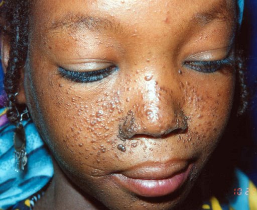 What is the probable underlying diagnosis for this case?
 Your Answer: Arteriovenous malformation
Correct Answer: Tuberous sclerosis
Explanation:Adenoma sebaceum is a type of skin lesion associated with tuberous sclerosis.
Tuberous sclerosis (TS) is a genetic condition that is inherited in an autosomal dominant manner. It is similar to neurofibromatosis in that most of the features seen in TS are neurocutaneous. The condition is characterized by various cutaneous features such as depigmented ‘ash-leaf’ spots that fluoresce under UV light, roughened patches of skin over the lumbar spine (Shagreen patches), adenoma sebaceum (angiofibromas) that are distributed like a butterfly over the nose, fibromata beneath nails (subungual fibromata), and café-au-lait spots. Neurological features include developmental delay, epilepsy (infantile spasms or partial), and intellectual impairment. Other features of TS include retinal hamartomas, rhabdomyomas of the heart, gliomatous changes that can occur in the brain lesions, polycystic kidneys, renal angiomyolipomata, and lymphangioleiomyomatosis, which is characterized by multiple lung cysts.
It is important to note that while café-au-lait spots are more commonly associated with neurofibromatosis, a study conducted in 1998 found that 28% of patients with TS also had café-au-lait spots. When comparing neurofibromatosis and TS, it is important to note that while they are both autosomal dominant neurocutaneous disorders, there is little overlap between the two conditions.
-
This question is part of the following fields:
- Dermatology
-
00
Correct
00
Incorrect
00
:
00
:
00
Session Time
00
:
00
Average Question Time (
Mins)